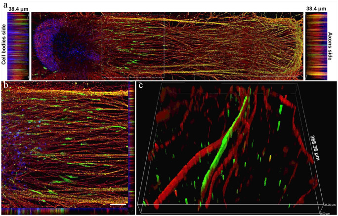
Figure 6.
Schwann cells migrated out of the spheroid and elongated along the axons. (a) Image showing how human Schwann cells (hSCs) stained for the hSC marker S100 (green) migrated out of the spheroid along with growing axons stained for βIII-tubulin (red) over a period of 4 weeks. Nuclei were labeled with DAPI (blue). Scale bar: 1000 µm. (b) High-magnification image of inset from image A. Scale bar: 25 µm. (c) 3D image showing close-up of the relationship between hSCs (green) and myelinated axons (red). Slice size was 368.36 × 368.36 × 34.00 µm. Adapted from Sharma et al.119

