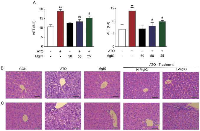
Figure 2.
Effect of MgIG on liver injury. (A) AST and ALT activity. Representative sections of hematoxylin-eosin staining in the hepatic (B) central vein and (C) duct area (magnification, ×400). Scale bar, 50 µm. CON showed normal hepatocyte architecture; ATO showed inflammation, hepatocyte vacuolation and necrosis/disorganization of the parenchyma. These symptoms decreased following MgIG treatment. Data are presented as the mean ± SEM (n=10). **P<0.01 vs. CON, #P<0.05 and ##P<0.01 vs. ATO. AST, aspartate aminotransferase; ALT, alanine aminotransferase; MgIG, magnesium isoglycyrrhizinate; CON, control; ATO, arsenic trioxide; H-, high; L-, low.

