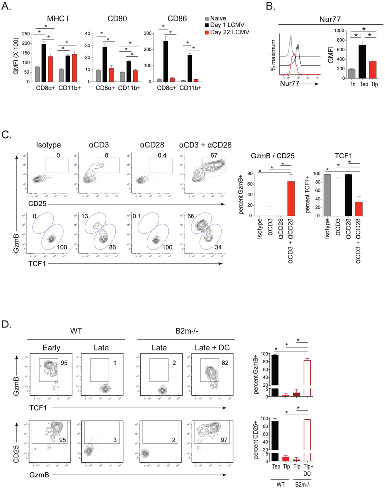
Figure 4. Diminished dendritic cell responsiveness and lower T cell stimulation/costimulation suppress differentiation of CD8+ effector Tlp cells while sustaining the TCF1+ subset.
(A) GMFI of MHC I, CD80 and CD86 on CD8α+ and CD11b+ DCs at 1 day (black) and 22 days post-Cl13 infection (red) (i.e., 1 day after early and late priming, respectively) and in naïve mice (gray).
(B) Nur77 expression by Tn, Tep, and Tlp P14 cells 60 hrs after priming.
C) P14 T cells were transferred into mice that had been infected 21 days earlier with Cl13. Four hours post-priming mice were treated i.p. with anti-CD3 and/or anti-CD28 or isotype control. Flow plots and bar graphs depict the frequency of GzmB+ CD25+ effector and TCF1+ memory-like P14 cells 60hrs after transfer.
(D) P14 T cells were transferred at the onset (Early) or at day 21 (Late) after Cl13 infection of wildtype or B2microglobulin (B2m)−/− mice. At the time of priming, a group of B2m−/− received GP33 peptide loaded bmDC. GzmB, TCF1 and CD25 expression was quantified on the P14 cells 60hrs after transfer.
Data represent 2-3 independent experiments with 4-5 mice per group. Error bars indicate SD. Significance was determined by one-way ANOVA. *, p<0.05.
See also Figure S4.

