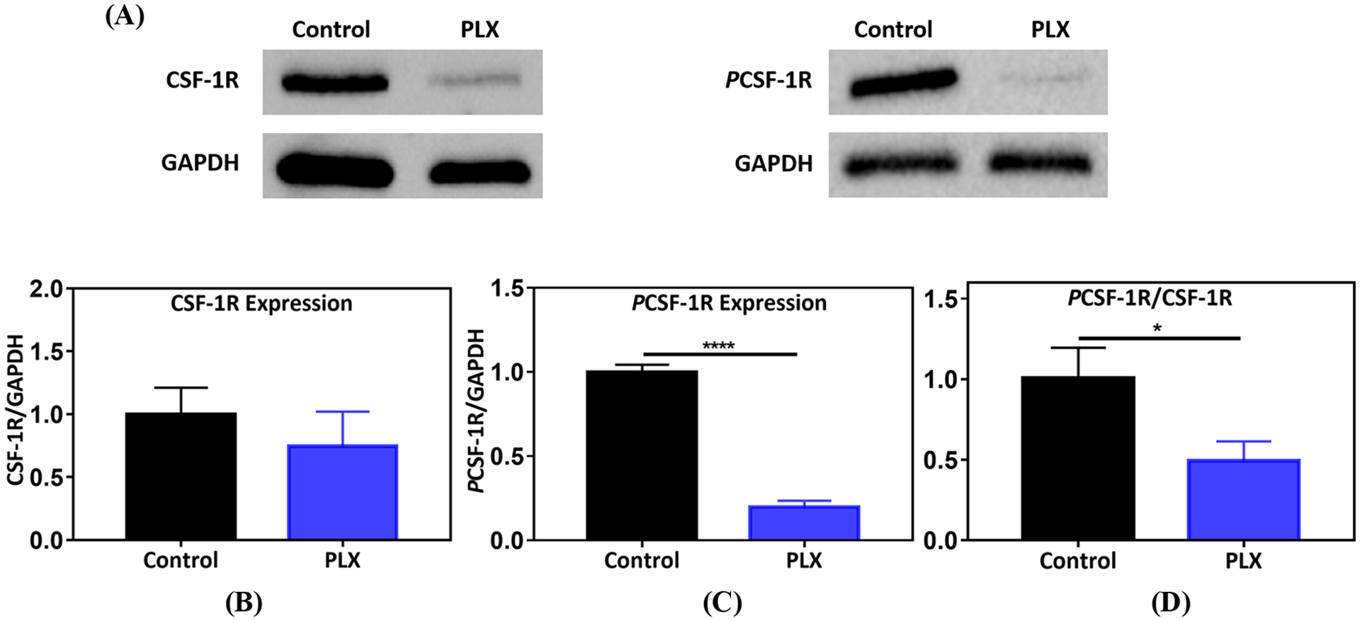
Figure 2.

Colony stimulating factor-1 receptor (CSF-1R) inhibition by PLX. (A) Protein bands for CSF-1R, PCSF-1R, and GAPDH, generated by western blot analysis on PLX (1 mg/kg, p.a.) treated lung tumor nodules, (B) effect of PLX on CSF-1R expression (n=7), (C) impact of treatment on PCSF-1R expression (n=7). For (B) and (C), protein densities were normalized to GAPDH, (D) ratio of PCSF-1R/CSF-1R. Control and PLX samples were normalized to average control in each independent experiment. Statistical significance was calculated by direct comparison using unpaired t test (*p <0.05,****p <0.0001), data represented as mean ±SEM.
