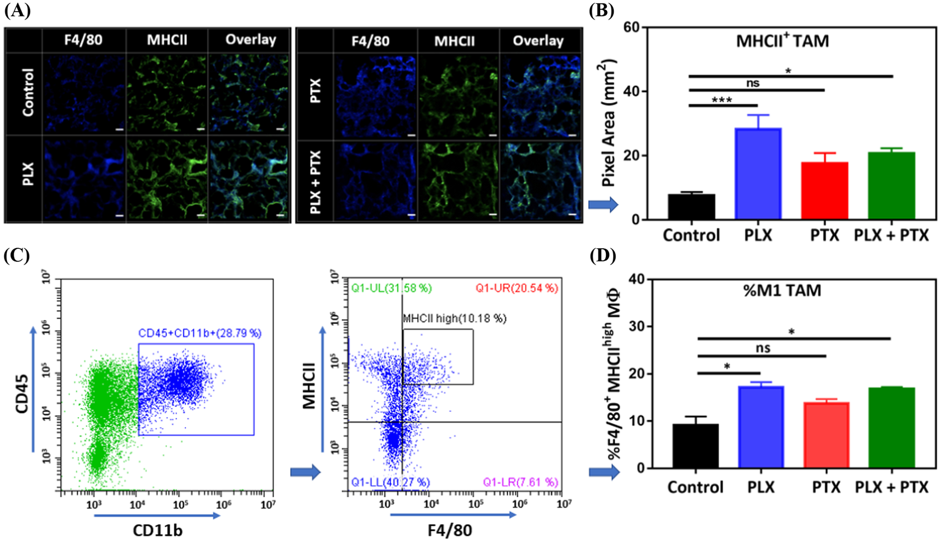
Figure 5.

PLX increases the population of MHCII+ TAM in the TME of 4T1 lung lesions. (A) A representative IF image acquired by confocal microscopy for total TAM (F4/80+), MHCII+, and MHCII+ TAM (F4/80+ MHCII+ overlay), (B) impact of PLX (p.a.) +/− PTX (i.v.), 1 mg/kg each, on MHCII+ TAM population, obtained from (A). Tumor nodules were collected from animal lungs (n=3) and six random images were taken on each group sample, pixels for MHCII+ TAM were converted to area (1pixel = 0.98 μm2) and plotted in (B). (C) Gating strategy on Flow-cytometry for tumor samples (control group), M1 TAM are represented by CD45+ CD11b+ F4/80+ MHCIIhigh cells, (D) effect of PLX (p.a.) +/− PTX (i.v.), 1 mg/kg each, on M1 TAM population obtained from (C) showing % of M1 TAM/CD45+ CD11b+. For (B) and (D), PLX +/− PTX treated groups were compared with control and statistical significance was calculated by One-way ANOVA (*p <0.05, ***p <0.001), data represented as mean ±SEM.
