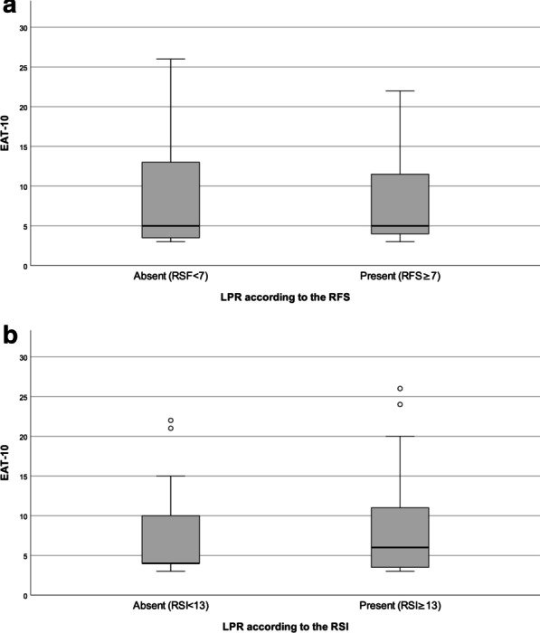
Fig. 2.

Comparison of the EAT-10 score between patients with and without LRP. In the box plots, vertical solid lines (whiskers) show lower and upper EAT-10 scores. Box stretches from lower hinge (25th percentile) to upper hinge (75th percentile). Median is shown as line across each box. Outsiders are represented by dots. a Comparison of the EAT-10 score between patients without LPR (n = 20, EAT-10 median 5, IQR 3.3–14) and patients with LPR (n = 15, EAT-10 median 5, IQR 3.8–13.3) according to the RFS (Mann–Whitney U test p = 0.934). b Comparison of the EAT-10 score between patients without LPR (n = 12, EAT-10 median 4, IQR 4–15) and patients with LPR (n = 23, EAT-10 median 6, IQR 3–11) according to the RSI (Mann–Whitney U test p = 0.717)
