
FIGURE 3.
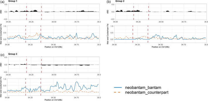
Distribution of the rIBD and F ST of the three groups (a–c) around the HMGA2 corresponding interval (34–35 Mb). The x‐axis displays the chromosomal coordinates, while the y‐axis shows the value of rIBD and F ST. In each group, the upper panel shows the estimation of rIBD. The positive value of rIBD suggests more similarity between neo‐bantam and bantam source than between neo‐bantam and normal‐sized counterpart; the negative rIBD value, on the contrary, shows the high haplotype sharing between neo‐bantam and normal‐sized counterpart. The lower panel displays the F ST estimation, the blue solid line represents the F ST between neo‐bantam and bantam source, while the orange dashed line displays F ST between neo‐bantam and normal‐sized counterpart