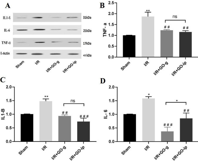
Figure 3.
GO attenuates I/R induced alterations in TNF α, IL 1β, and IL 6 expression levels. Representative images of Western blot results for inflammatory markers (A). The levels of TNF-α (B), IL-1β (C), and IL-6(D) in renal tissues of rats. * P<0.05 and ** P<0.01 compared with sham control. ## P<0.01 and ###P<0.001 compared with I/R. ns: non-significant. All values are mean±SEM

