Figure 1.
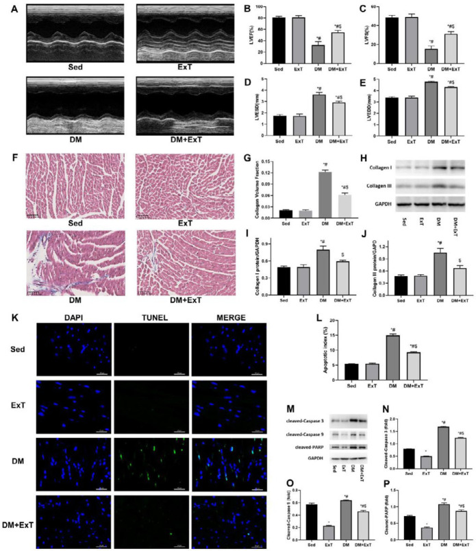
Exercise alleviates cardiac remodelling in mice with DCM. (A) Representative transthoracic M-mode echocardiographs. (B-E) Measurements of LVEF, LVFS, LVESD and LVEDD (n=5). (F) Representative images of Masson’s trichrome staining showing collagen deposition. (G) Quantification of collagen volume fraction using Image-Pro Plus (n=5). (H-J) Representative immunoblots and semiquantitative analysis of collagen I and collagen III. (K) Representative immunofluorescent images of staining with DAPI (blue) and TUNEL (green). (L) Quantitative assessments of apoptotic indexes (n=5). (M-P) Representative immunoblots and semiquantitative analysis of cleaved-caspase 3, cleaved-caspase 9, and cleaved-PARP. Sed, sedentary C57BL/6 mice; ExT, exercised C57BL/6 mice; DM, C57BL/6 mice with diabetes; DM+ExT, diabetic C57BL/6 mice with exercise intervention; LVEF, left ventricular ejection fraction; LVFS, left ventricular fractional shortening; LVESD, left ventricular end-systolic internal dimension; LVEDD, left ventricular end-diastolic internal dimension. The data are presented as the means±SEM of triplicate experiments, and group differences were determined by one-way ANOVA followed by Tukey’s multiple comparisons test. *, P<0.05 vs Sed; #, P<0.05 vs ExT; $, P<0.05 vs DM

