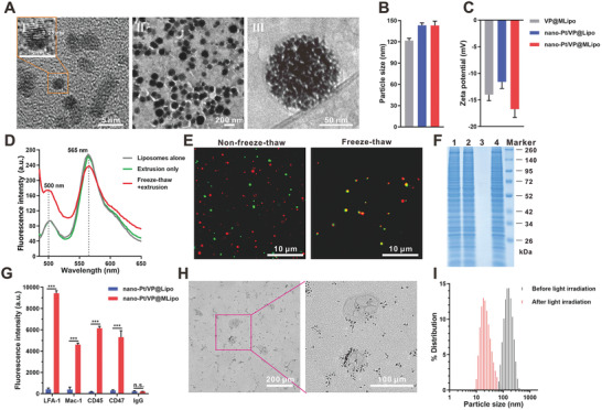
Figure 1.

Characterizations of nano‐Pt/VP@MLipo. A) TEM images of free I) nano‐Pt and II) nano‐Pt/VP@MLipo. III) An enlarged Cryo‐TEM image of nano‐Pt/VP@MLipo. B) Particle size and C) zeta potential measured through dynamic light scattering (DLS). D) Liposomes were labeled with DiO‐DiI FRET pair. The recovered fluorescence of DiO at ≈500 nm and meanwhile weaken FRET signal at 565 nm indicated the fusion of liposomes and Mφ CM vesicles. E) CLSM images of the mixture of Mφ CM vesicles (DiO, green) and liposomes (DiR, red) subjected to freeze–thaw process or not. F) SDS‐PAGE of the samples with equivalent protein contents (25 µg). 1) Mφ CM; 2) MLipo (empty); 3) nano‐Pt/VP@Lipo; 4) nano‐Pt/VP@MLipo. G) Flow cytometry validation of the functional proteins (LFA‐1, Mac‐1, CD45, and CD47) and their orientation on the liposome surface. H) TEM observation of the light (690 nm) irradiation‐triggered nano‐Pt liberation from the aqueous cavity of nano‐Pt/VP@MLipo. I) Light irradiation‐induced size change identified using DLS. Data are presented as mean ± s.d. (n = 3). ***p < 0.001.
