
Figure 3.
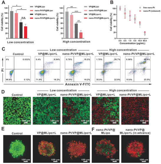
Cytotoxicity of nano‐Pt/VP@MLipo with light (L) irradiation or nano‐Pt alone in 4T1 cells and tumor spheroids. A) The cells were incubated with liposomes for 4 h, and irradiated with 690 nm laser (100 mW cm−2) for 1 min. Cell viabilities were examined using CCK‐8 after additional 24 h incubation in fresh medium. B) Cell viability after treatment with free or released nano‐Pt for 48 h. The released nano‐Pt was obtained by light irradiation on the nano‐Pt/VP@MLipo for 1 min. C) Cell apoptosis induced by liposomes plus light irradiation was examined via flow cytometry. The cells were stained with Annexin V‐FITC and PI. D) 4T1 cells treated with liposomes plus irradiation were stained with calcein‐AM and PI for viable (green) and dead (red) cells imaging, respectively. E) Cytotoxicity of liposomes with/without light irradiation in 4T1 tumor spheroids. F) Cytotoxicity of intact liposomes (containing 50 µg mL−1 Pt) or those previously treated by light irradiation in 4T tumor spheroids. Low (50 × 10−9 m VP, 0.715 µg mL−1) and high (200 × 10−9 m VP, 2.86 µg mL−1 Pt) concentrations of liposomes were used in (A), (C), and (D), and high concentration in (E). Data are presented as mean ± s.d. (n = 3). *p < 0.05, ***p < 0.01, and ***p < 0.001.