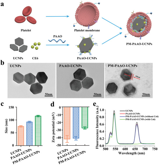
Figure 1.

Design and characterization of PM‐PAAO‐UCNPs. a) Schematic illustration showing the composition of PM‐PAAO‐UCNPs. b) Transmission electron microscopy images of UCNPs, UCNPs loaded in micelles (PAAO‐UCNPs), and platelet member‐coated PAAO‐UCNPs (PM‐PAAO‐UCNPs). c) Size and d) zeta potential of UCNPs, PAAO‐UCNPs, and PM‐PAAO‐UCNPs in PBS. Data are presented as mean ± SD (n = 3). e) Fluorescence spectra of UCNPs, PAAO‐UCNPs, and PM‐PAAO‐UCNPs with or without Ce6 under 980 nm laser irradiation.
