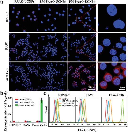
Figure 2.

Binding ability of PM‐PAAO‐UCNPs to foam cells in vitro. a) Confocal laser fluorescence imaging of RAW, HUVEC, and foam cells incubated with PAAO‐UCNPs, EM‐PAAO‐UCNPs, and PM‐PAAO‐UCNPs, respectively. Blue and red fluorescence indicates nuclei and UCNPs, respectively. b) Quantification on the basis of Er concentration in cells. Data are presented as mean ± SD (n = 3). c) Flow cytometry analysis of respective affinity of PAAO‐UCNPs, EM‐PAAO‐UCNPs, and PM‐PAAO‐UCNPs to RAW, HUVEC, and foam cells.
