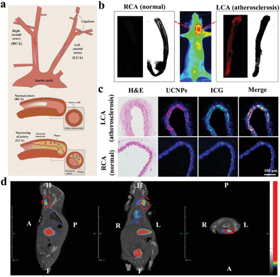
Figure 3.

Targeting ability of PM‐PAAO‐UCNPs to atherosclerotic plaque in vivo. a) Schematic illustration of partial carotid ligation surgery model. The left carotid artery (LCA) was ligated and the right carotid artery (RCA) was used as control. b) Fluorescence images of mice injected with PM‐PAAO‐UCNPs (middle) and isolated arteries (left and right). c) H&E staining and fluorescent imaging of artery sections. Nuclei: blue; UCNPs: red; ICG: green; merge of ICG and UCNPs: yellow. d) SPECT/CT images of PM‐PAAO‐UCNPs (labeled with 125I) including transverse, coronal, and sagittal sections in model mouse 1 h following injection. Plaque region is denoted by the red circle.
