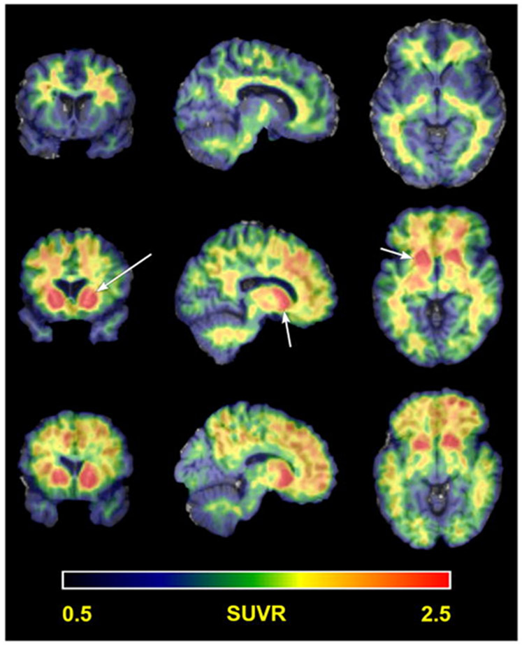
Figure 3 ∣. Amyloid PET in Down syndrome.
Representative examples of Pittsburgh Compound B (PiB) PET neuroimaging for amyloid in people with DS. The pattern of PiB binding highlights striatal PiB uptake (arrows). Three patterns of PiB update are highlighted in this figure from Lao and colleagues (2016)86 showing nonspecific white matter binding in the top row, striatal only PiB uptake in the middle row and striatal with cortical PiB update on the bottom row. SUVR, standard uptake value ratio.

