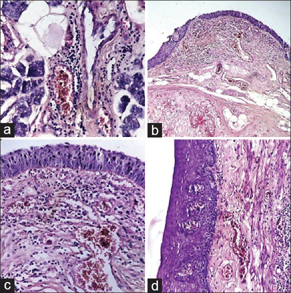
Figure 5.

(a) Submandibular salivary gland: Perivascular lymphocytic infiltrate. (H and E, ×400). (b) Epiglottis: Subepithelial lymphocytic infiltrate and congestion (H and E, ×100). (c) Epiglottis: Subepithelial lymphocytic infiltrate and congestion (H and E, ×400). (d) Esophagus: Subepithelial congestion and lymphocytic infiltrate (right side of the picture) (H and E, ×200)
