
Figure 2.
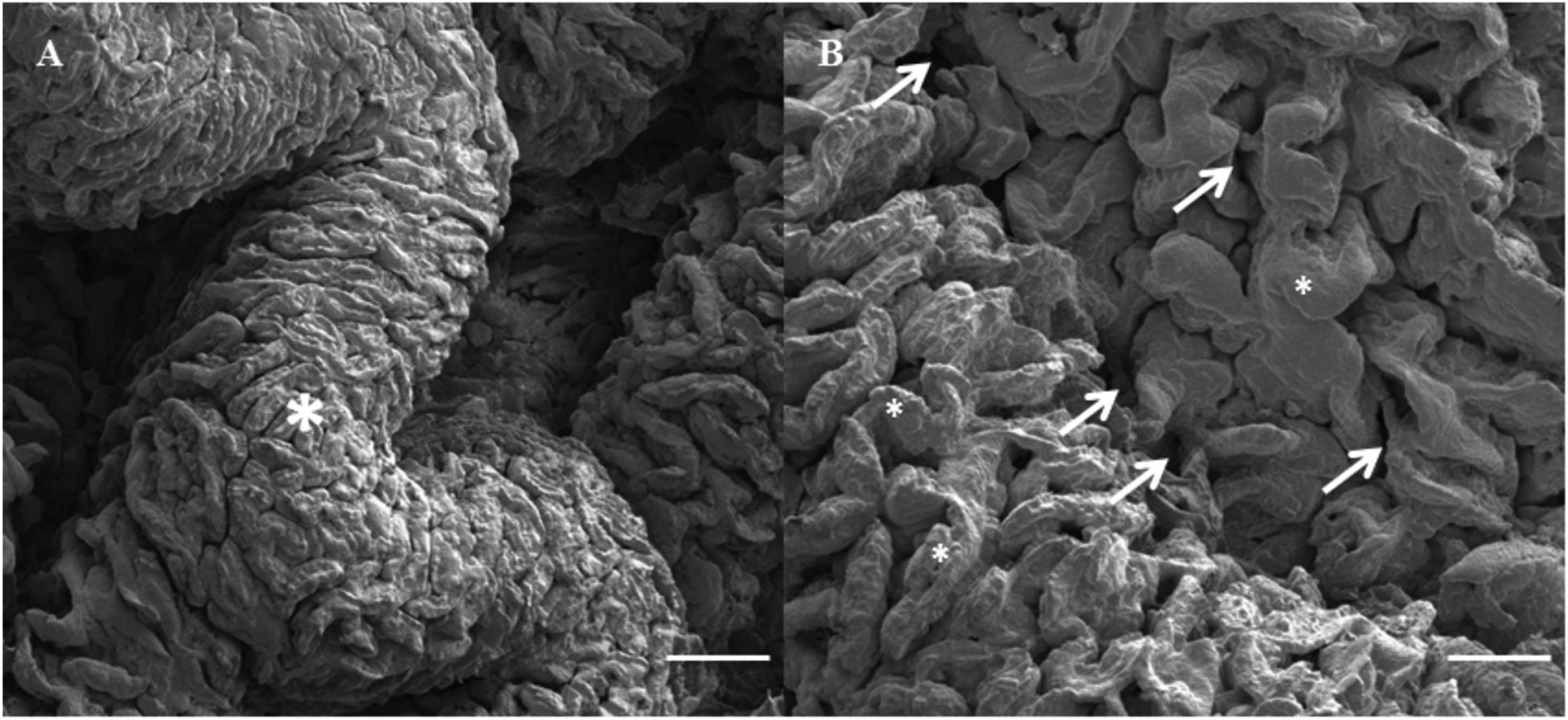
SEM images show varying topographical features of osmium fixed native small intestinal tissue, including (a) macroscopic folds (*) and (b) microscopic villi (*) and crypts (arrows). Bar = 1 mm in (a) and 500 μM in (b).
Official websites use .gov
A
.gov website belongs to an official
government organization in the United States.
Secure .gov websites use HTTPS
A lock (
) or https:// means you've safely
connected to the .gov website. Share sensitive
information only on official, secure websites.
SEM images show varying topographical features of osmium fixed native small intestinal tissue, including (a) macroscopic folds (*) and (b) microscopic villi (*) and crypts (arrows). Bar = 1 mm in (a) and 500 μM in (b).