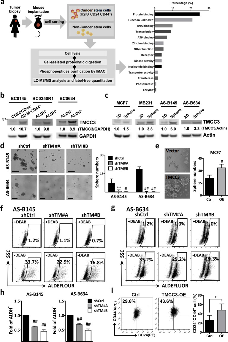
Fig. 1. TMCC3 is highly expressed in BCSCs and crucial for BCSC maintenance.
a Workflow illustration of phosphoproteomic analysis using BCSCs and non-BCSCs of BC0145 PDX tumor to discover function unknown phosphoproteins in BCSCs. b The protein levels of TMCC3 in BCSCs and non-BCSCs FACS-sorted from breast cancer PDX tumors, BC0145, BC0350R1, and BC0634 using H2kd-CD24−CD44+ (BC0145) and H2kd−ALDH+ (BC0350R1 and BC0634), respectively. c The protein levels of TMCC3 in MCF7, MDA-MB231 (MB231), AS-B145, and AS-B634 cultured as monolayer (2D) and mammosphere culture (Sphere) condition, as determined by western blotting. d The representative images of mammosphere in TMCC3 silenced AS-B145 and AS-B634 (left panels). Sphere forming capacities of AS-B145 and AS-B634 transduced with shRNA-TMCC3 (shTM #A and shTM #B) vs shRNA-control (shCtrl) after 7 days culture (right panels). Data represent mean ± SD. **p < 0.01, #p < 0.001 and ##p < 0.0001 (n = 6, t-test). Scale bar = 200 μm. e The representative images of mammospheres and sphere forming capacities of MCF7 overexpressing TMCC3 vs vector control after 7 days culture. Data represent mean ± SD. #p < 0.001 (n = 6, t-test). Scale bar = 200 μm. f, g ALDH activity of TMCC3 silenced AS-B145 and AS-B634 (shTM #A and shTM #B), as compared with their respective controls. ALDH+ subpopulation was determined by flow cytometry and the relative folds of ALDH+ cells were calculated from three independent experiments (h). Data represent mean ± SEM. ##p < 0.0001 (n = 3, t-test). i CD24−CD44+ cell population of TMCC3-overexpressed MCF7 was determined by flow cytometry, as compared with vector control transduced MCF7. Data represent mean ± SEM. *p = 0.01 (n = 6, t-test).

