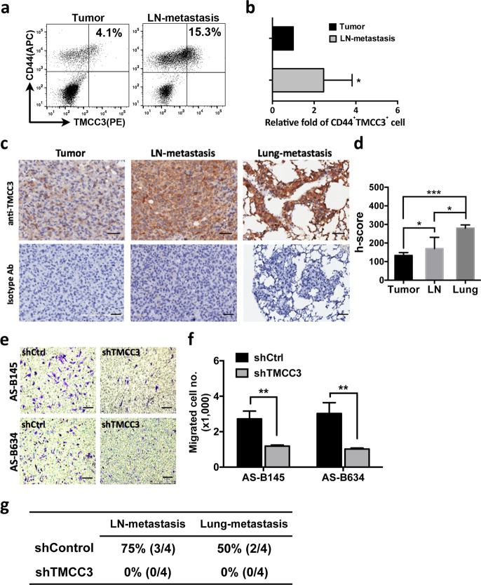
Fig. 3. TMCC3 contributes to tumor metastasis in vitro and in vivo.
a FACS analysis of TMCC3 expression in permeabilized BCSC (CD44+) harvested from primary tumor (Tumor) and metastatic lymph node (LN-metastasis) of BC0145 PDX tumor. b Percent of CD44+TMCC3+ cell population in metastatic lymph node was normalized to that in primary tumor to show the relative folds. *p < 0.05 (n = 5, t-test). c The representative IHC stainings of primary tumor, lymph node, and lung-metastatic tissues from one of three BC0634 bearing mice. Scale bar = 100 μm. d The histoscores (h-score) of TMCC3 expression in tumors of three mice were calculated. *p < 0.05, ***p < 0.001 (n = 3, t-test). e, f The numbers of migrated cells in each group were determined in trans-well migration assay. **p < 0.01 (n = 3, t-test). Scale bar = 100 μm. g Metastatic frequency of TMCC3 silenced AS-B634. 2 × 10⁴ shTMCC3 or shControl transduced AS-B634 were implanted into mammary fat pad of NSG mice (n = 4). Tumor, lymph node, and lung of tumor bearing mice were harvested at 3 months after implantation.

