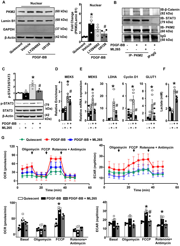
Figure 5. PKM2 activator inhibits STAT3, and β catenin induced transcription of MEK5 and LDHA in human coronary artery SMC.
A, HCaSMCs were pretreated with Akt inhibitor (LY294002, 10 μM) or ERK pathway inhibitor (U0126, 10 μM) and stimulated with PDGF-BB (20 ng/ml) for 6 h. PKM2, Lamin B1, GAPDH, and β-actin were detected in the nuclear extract by immunoblotting. The left panels show representative immunoblots, and the right panel shows the densitometric analysis for PKM2/Lamin B1 (n = 4/group). B, HCaSMCs were stimulated with PDGF-BB (20 ng/ml) for 6 h, and cell extracts were immunoprecipitated (IP) with PKM2 antibody or control IgG and immunoblotted for PKM2, STAT3, and β-catenin. C, HCaSMCs were pretreated with ML265 (50 μM) and stimulated with PDGF-BB (20 ng/ml) for 6 h. Representative immunoblots and densitometric analysis for p-STAT3, total STAT3, and β-actin (n = 6/group). D, The ChIP assay of the MEK5 promoter in PDGF-BB stimulated HCaSMCs using antibodies against PKM2. ChIP using rabbit Ig as a negative control (n = 6/group). E, Real-time quantitative PCR analysis of MEK5, LDHA, Cyclin D1, and GLUT1 (n = 5/group). The results are presented as changes in PCR products normalized to GAPDH. (F), The bar graph shows lactate levels in cell supernatant (n = 4/group). (G), HCaSMCs were seeded in 96 well XF cell culture microplate. OCR and ECAR were measured in response to PDGF-BB in the presence or absence of ML265 (50 μM) (n =4). Values are mean ± SEM. Statistical analysis: A, C and F, One-way ANOVA followed by Bonferroni’s post hoc test; E, Kruskal-Wallis test (non-parametric One-way ANOVA) followed by uncorrected Dunn’s test; G, two-way ANOVA followed by uncorrected Fisher's LSD test. *P < 0.05 vs. quiescent; #P<0.05 vs. PDGF-BB-stimulated cells.

