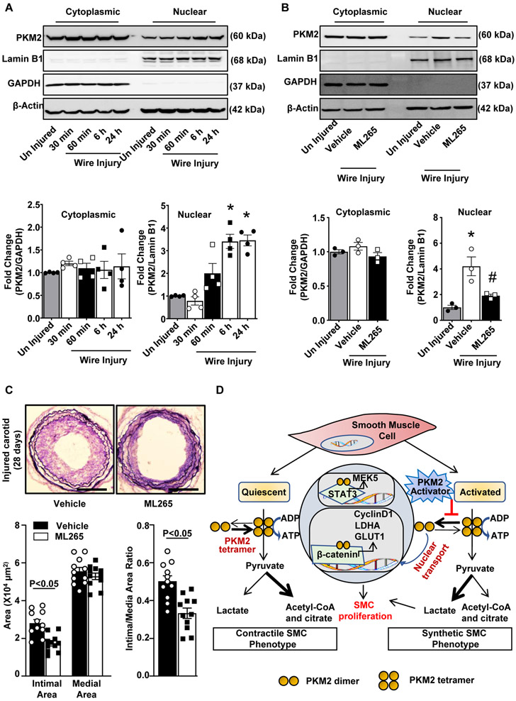
Figure 6. PKM2 activator inhibits injury-induced neointimal hyperplasia in wild-type mice.
A, Wire injury was done in the carotid artery of wild-type mice. Cytosolic and nuclear extracts were prepared at the following time points: 30 min, 60 min, 6 h, and 24 h after wire injury and then subjected to immunoblotting of PKM2, Lamin B1, GAPDH, and β-actin. The lower panel shows the densitometric analysis for PKM2/GAPDH in cytosolic extract and PKM2/Lamin B1 in nuclear extract (n = 4/group). B, Wire injury was done in the carotid artery of wild-type mice pretreated with ML265 (50 μM). Cytosolic and nuclear extracts were prepared 6 h after wire injury and were subjected to Western blotting of PKM2, Lamin B1, GAPDH, and β-actin. The lower panel shows the densitometric analysis for PKM2/GAPDH in cytosolic extract and PKM2/Lamin B1 in nuclear extract (n = 3/group). C, Carotid arteries pretreated with ML265 (50 μM in 30% w/v pluronic gel) were analyzed 28 days after wire injury. The left panel shows representative images of Verhoeff’s van Gieson stained carotid artery sections. Scale bars: 200 μm. The right panels show quantification of intimal area, medial area, and a ratio of intimal to the medial area. (n = 11/group). Each dot represents a single mouse. D, Schematic showing the mechanism by which PKM2 mediates SMC proliferation and neointimal hyperplasia. Values are expressed as mean ± SEM. Statistical analysis: A, B, 1-way ANOVA with Bonferroni’s post hoc test; C, unpaired Student’s t-test. *P < 0.05 vs. uninjured; #P<0.05 vs. wire injury + vehicle.

