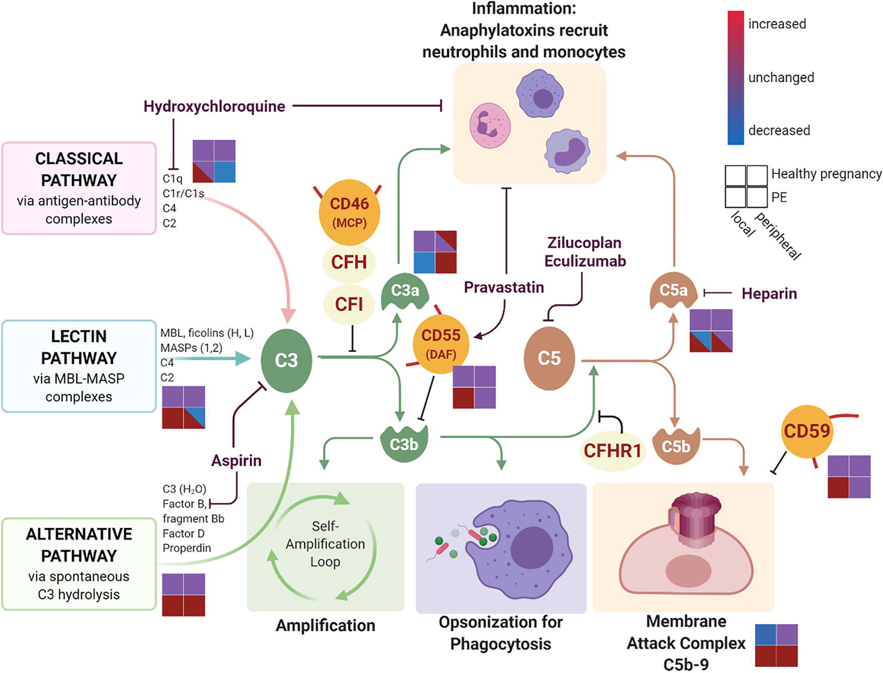
Figure 1. Local and systemic complement activation in preeclampsia and healthy pregnancy and complement-modulating medications.

The complement cascade is initiated through the classical, lectin, and alternative pathways. Complement activation results in inflammation primarily mediated through anaphylatoxins C3a and C5a, pathogen opsonization for phagocytosis via C3b, and cell lysis through the terminal membrane attack complex formation. Endogenous soluble regulators of complement activation (CFH, CFI, CFHR1) and membrane-bound regulators (MCP, DAF, and CD59) work through inhibition of the complement pathway at different steps. Various pharmacologic agents also target the complement activation at different steps. Schematic for the level of complement component activation is represented for healthy pregnancy (upper two squares) and in preeclampsia (PE, lower two squares) and separated by local decidua complement activation (left squares) and peripheral complement activation (right squares). Complement component levels are increased (red), decreased (blue), or unchanged (purple) depending on the disease state and location in pregnancy. Created with BioRender.com
