Figure 1.
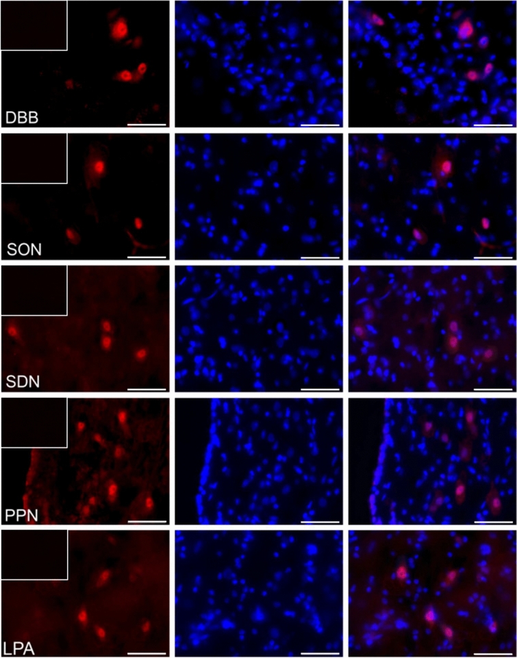
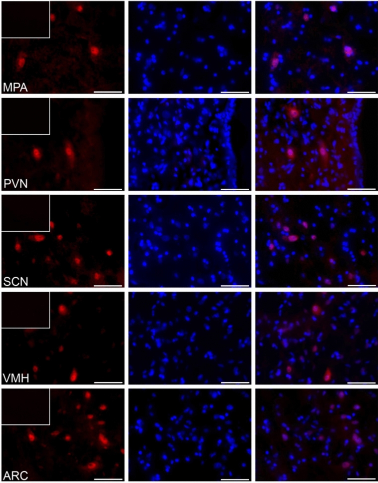
The visfatin localization in the porcine hypothalamus on days 10–12 of the estrous cycle. Immunoreactivity of visfatin was determined by fluorescent immunohistochemistry. Columns from left: first—visfatin expression, visualized by Alexa Fluor 555 as red fluorescence; second—nuclei stained with DAPI, visualized as blue fluorescence; third—merged images of channels (Olympus BX51, Olympus Soft Imaging Solutions, Germany). In the upper left corner of each image in the first column—negative control, where the primary antibody were replaced with non-immunosera. Particular images indicate the immunolocalization of visfatin in the diagonal band of Broca (DBB), supraoptic nucleus (SON), preoptic area (SDN), periventricular nucleus (PPN), lateral (LPA) and medial preoptic area (MPA), paraventricular nucleus (PVN), suprachiasmatic nucleus (SCN), arcuate nucleus (ARC), as well as ventromedial nucleus (VMH). Immunofluorescent staining was repeated on three pigs. Scale bar: 50 μm.


