Figure 2.
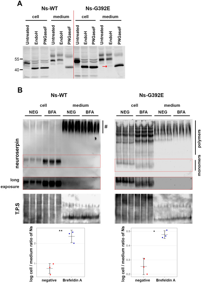
G392E-mutant neuroserpin is secreted via the ER-to-Golgi secretory pathway. (A) Cell extracts (cell) and culture media (medium) from HEK-293 cells overexpressing wild-type or G392E-mutant neuroserpin were collected and treated with EndoH or PNGaseF to remove N-linked mannose-rich or all types of N-linked oligosaccharides, respectively. In the cell extracts, both wild-type and mutant neuroserpin were completely deglycosylated by treatment with both enzymes. In the media, however, whereas wild-type neuroserpin acquired EndoH resistance, a faint band of Endo H-sensitive mutant neuroserpin could be detected (red arrow). The result shown is representative of three independent experiments. (B) HEK-293 cells stably transfected with wild-type or G392E-mutant neuroserpin were treated with BFA or solvent alone (NEG) for 4 h. Cell extracts (cell) and culture media (medium) were collected and analyzed by non-denaturing PAGE and western blot using an antibody directed against neuroserpin. As loading control, all proteins were stained (total protein stain, T.P.S). For wild-type neuroserpin, strong intracellular accumulation was observed upon treatment. Since culture medium was collected after a short incubation of 4 h, reduced secretion of monomeric neuroserpin was visible only after long exposure of the membrane (middle panel). Smears visible in the medium of wild-type neuroserpin-transfected cells (#) represent background signal, as this was also observed in cells transfected with vector only (see Fig. 1b). Similarly, mutant neuroserpin was increasingly retained within the cells treated with BFA, leading to enhanced polymerization, and less polymeric forms were detected in the media. For quantification, the mean ratio of the normalized density data (cell/medium) was compared. n = 3; p = 0.0027 for wild-type; p = 0.0104 for G392E-mutant. Three independent experiments with three technical replicates each were performed and a representative one is shown. Western blot membranes shown in (a) and (b) have been cropped, full-length blots are presented in Supplementary Fig. S3.

