Figure 3.
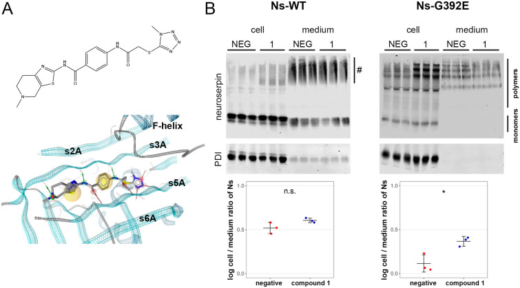
Secretion of G392E-neuroserpin can be pharmacologically regulated. (A) 2D structure (top) of compound 1 and visualization of its predicted binding mode (bottom). The protein backbone of neuroserpin is visualized as a ribbon. Green and red arrows indicate hydrogen bonds predicted to be formed between the ligand and the protein, and yellow spheres mark areas in the compound predicted to engage in hydrophobic interactions. Visualization with LigandScout37. (B) HEK-293 cells stably transfected with wild-type or G392E-neuroserpin were treated with the small organic compound 1 or solvent alone (NEG) for 18 h. Cell extracts (cell) and culture media (medium) were collected and analyzed by non-denaturing PAGE followed by western blot using an anti-neuroserpin antibody. PDI was used as loading control. Smears visible in the medium of wild-type neuroserpin-transfected cells (#) represent background signal, as they were also observed in cells transfected with vector only (see Fig. 1b). For quantification, the mean ratio of the normalized density data (cell/medium) was compared. n = 3; p = 0.1292 for wild-type; p = 0.0277 for G392E-mutant. Two independent experiments with three technical replicates each were performed and a representative one is show. Western blot membranes shown in (B) have been cropped, full-length blots are presented in Supplementary Fig. S3.

