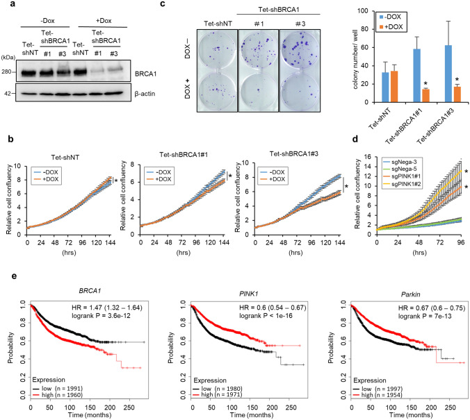
Figure 5.
BRCA1 knockdown represses breast cancer cell growth and lower BRCA1 expression correlates with longer relapse-free survival rate. (a) MCF7 cells stably expressing Tet-shRNA vector against BRCA1 (Tet-shBRCA1) or control vector (Tet-shNT) were cultured with or without 0.1 μg/ml DOX for 48 h. BRCA1 knockdown efficiency was confirmed by Western blotting. (b) Growth of MCF7 cells expressing the Tet-shNT, Tet-shBRCA1 #1, or #3 vectors was assessed using the IncuCyte live-cell imaging system in the presence or absence of 0.1 μg/ml DOX for 4 days. n = 4, means ± S.D. *p < 0.05 versus -DOX. (c) Comparison of colony formation ability between MCF7 cells expressing Tet-shNT and Tet-shBRCA1. Cells were seeded in 6-well plates and cultured for 3 weeks in the presence or absence of 0.1 μg/ml DOX. Visualized colonies are shown in the left panel. Summarized colony numbers are shown in the right panel. Bar = means ± S.D. n = 3, *p < 0.05 versus -DOX. (d) The growth of PINK1 knockout MCF7 cells (sgPINK1#1, #2) or control cells (sgNega3, 5) was assessed using the IncuCyte live-cell imaging system. n = 5, means ± S.D. *p < 0.05 versus sgNega5. (e) Relapse-free survival rates of breast cancer patients were compared between high and low BRCA1, Parkin, or PINK1 expression in patients. Data are shown as Kaplan–Meier plots; graphs were generated using an online Kaplan–Meier plotter.

