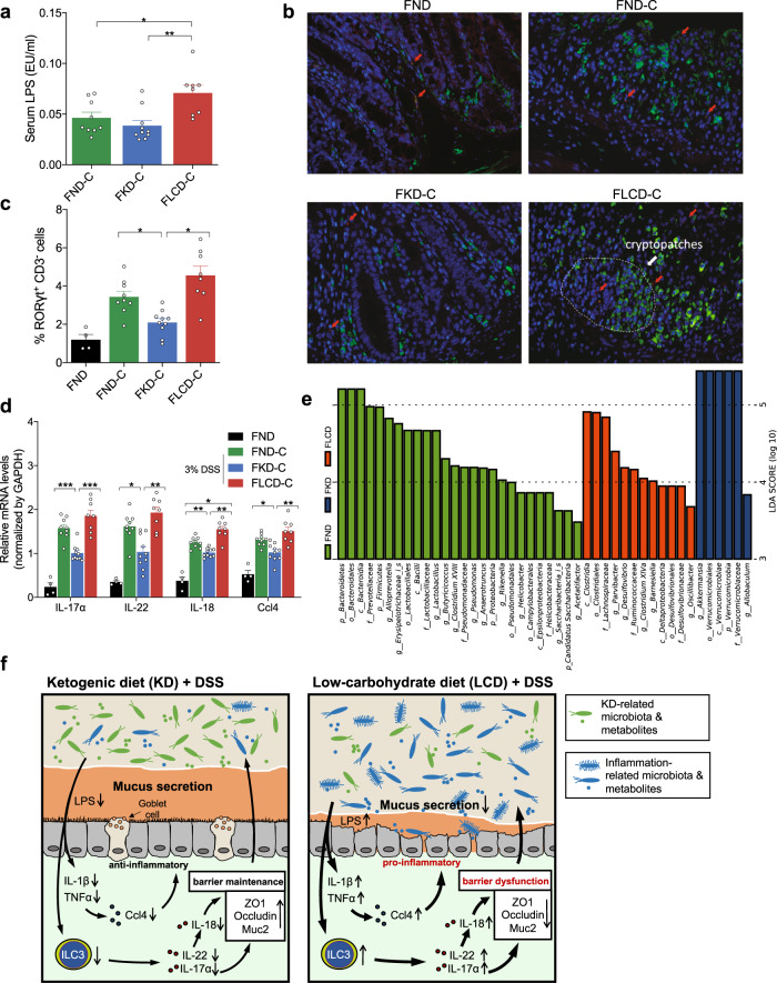
Fig. 7.
Effects of FMT from KD- and LCD-fed mice on mucosal inflammation and immunity in colitis-induced germ-free mice. a Serum LPS levels detected by end-point chromogenic tachypleus amebocyte lysate in DSS-induced colitis germ-free mice after FMT from mice fed with KD (FKD-C), LCD (FLCD-C), or ND (FND-C). b Representative immunofluorescence images of RORγt+CD3− ILC3 cells in colon tissues (original magnification ×400). Green: positive staining of RORγt; Red arrows: positive staining of CD3. c Percentage of RORγt+CD3− ILC3 cells in colonic sections. d Quantitative RT-PCR confirming changes in IL-17α, IL-18, IL-22, and Ccl4 expression. e Histogram of linear discriminant analysis scores for differentially abundant bacteria in each group (p: Phylum, c: Class, o- Order, f: Family, g: Genus). f Schematic summary for the proposed role of KD (left) and LCD (right) in colitis. *p < 0.05; **p < 0.01; ***p < 0.001. (FKD-C, n = 10, FLCD-C, n = 8; FND-C, n = 9; FND, n = 4). Results are expressed as mean ± SEM (a, c, d). KD ketogenic diet, LCD low-carbohydrate diet, ND normal diet, DSS dextran sulfate sodium, OTU operational taxonomic units, PCoA principal component analysis, ILC3 group 3 innate lymphoid cells

