Figure 5.
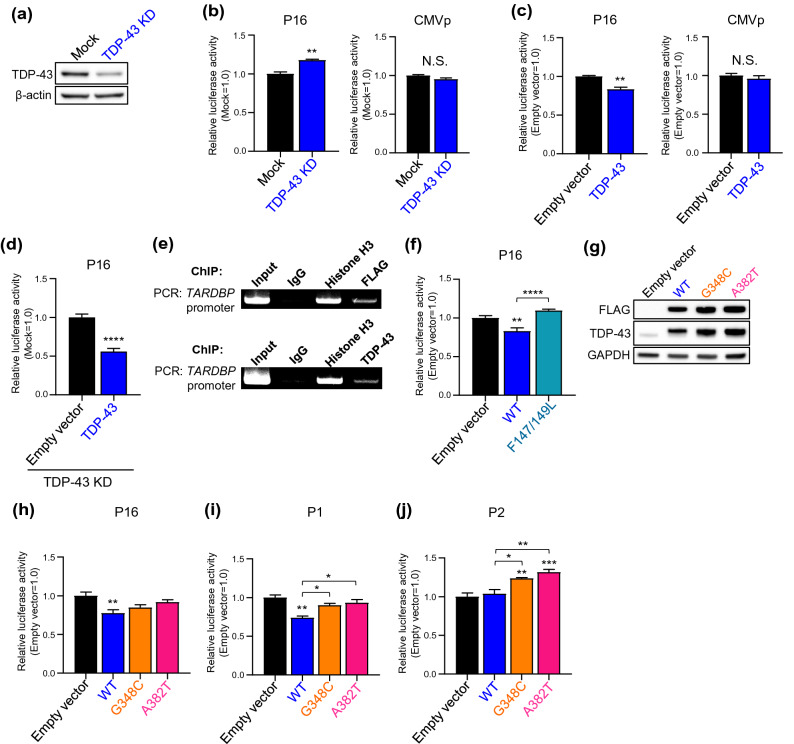
TDP-43 regulates the activity of the TARDBP upstream and intron 1 promoter. (a) Immunoblot analysis of TDP-43 in HeLaS3 cells. Mock and KD indicate transfected negative control or TDP-43 siRNA, respectively. (b-d) Luciferase assay of P16 or CMV promoter activity with TDP-43 KD (b) or overexpression (c) or both endogenous TDP-43 KD and exogenous TDP-43 overexpression (d). n = 4 (b, c) and n = 8 (d) replicates; Welch’s t-test relative to Mock (b) or Empty vector (c,d). (e) ChIP assay of overexpressed FLAG-TDP-43 (top) or endogenous TDP-43 (bottom) binding to the TARDBP promoter in HEK293T cells. Immunoprecipitation was performed using anti-DDDDK (FLAG) antibody (for overexpressed FLAG-TDP-43) or anti-TDP-43 antibody (for endogenous TDP-43), then PCR was conducted using primer detecting TARDBP -721 to -431 (290 bp). Control IgG and Histone H3 antibody are used for negative or positive control for immunoprecipitation. (f) Luciferase assay of P16 promoter activity with RRM1 mutant TDP-43 (F147/149L) overexpression. n = 8 replicates; one-way ANOVA with post hoc Tukey’s test. (g) Immunoblot analysis of FLAG-WT or 2 pathological TDP-43 mutants (G348C or A382T) overexpressed in HeLaS3 cells. Empty vector was transfected as the negative control. (h-j) Luciferase assay of P16 (h), P1 (upstream promoter) (i) and P2 (intron 1 promoter) (j) promoter activity with overexpression of WT TDP-43 or 2 pathological TDP-43 mutants (G348C or A382T). n = 7 (h), n = 3 (i) and n = 5 (j) replicates; one-way ANOVA with post hoc Tukey’s test. 2 μg of each reporter plasmids (P16, P1 and P2) were co-transfected with 1 μg of TDP-43/pFLAG-CMV2 in all overexpression experiments. Empty vector was used as the control in the overexpression experiment. All error bars indicate the means ± SEMs. *P < 0.05, **P < 0.01, ***P < 0.001, ****P < 0.0001.

