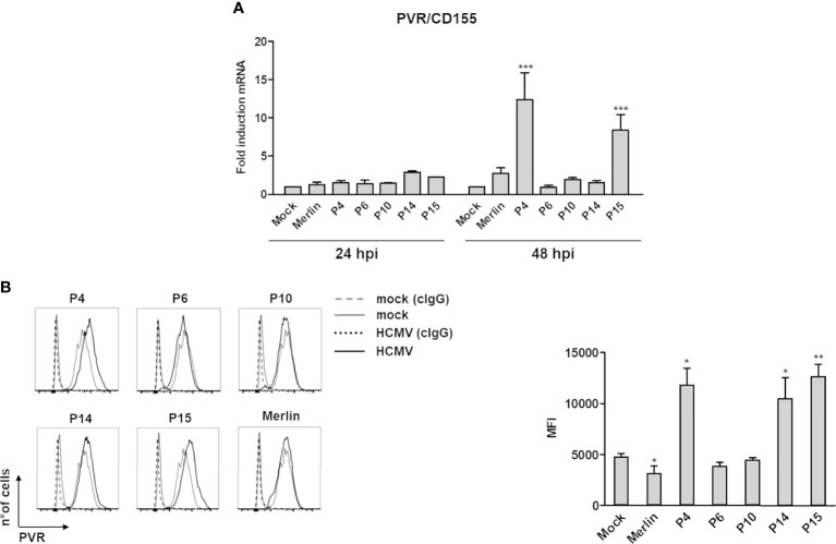
Figure 1.
Modulation of the NK cell ligand PVR/CD155 by HCMV clinical isolates. (A) Primary human foreskin fibroblasts (HFFs) infected with the indicated clinical isolates (P4, 6, 10, 14, and 15), the Merlin strain, or uninfected (mock) were co-cultured with an excess of uninfected HFFs, as described in Materials and Methods, and subjected to RT-qPCR to measure PVR/CD155mRNA expression levels. Values were normalized to the housekeeping gene glyceraldehyde-3-phosphate dehydrogenase (GAPDH) mRNA and plotted as fold induction relative to mock-infected cells (set at 1). Data from three experiments performed at 24 and 48 hours post-infection (hpi) are shown. Error bars show standard deviation (SD) (***P < 0.001; two-way ANOVA followed by Bonferroni’s post-tests, for comparison of infected vs. mock cells). (B) FACS analysis assessing PVR/CD155 expression at 3 days post-infection. Left panel: a representative experiment of at least four performed with all HCMV isolates is shown. Dashed and dotted lines indicate isotype control in mock or HCMV-infected cells, respectively. Right panel: data derived from at least four experiments performed with all HCMV isolates. PVR/CD155 expression levels are presented as mean fluorescence intensity (MFI) ± SE (*P < 0.05; **P < 0.01, paired Student t test for comparison of infected vs. mock cells).

