Figure 2.
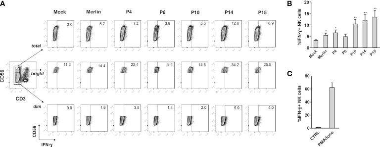
IFN-γ expression in NK cells co-cultured with HCMV-infected HFFs. NK cells were co-cultured with mock- or HCMV-infected HFFs at day 2 post infection, as described in Materials and Methods. The day after, NK cells were harvested and stained for intracellular IFN-γ. (A) A representative experiment of at least four performed with all HCMV isolates is shown. Numbers indicate the percentage of IFN-γ+ cells in the gate of CD3-CD56+ (total), CD3-CD56dim (dim), or CD3-CD56bright (bright) NK cells. All cells were first gated among viable (Zombie-) population. (B) Cells were analyzed as in panel (A), and data are expressed as the mean percentage (%) ± SE of IFN-γ+ cells in the gate of total CD3-CD56+ NK cells. Data are from at least four independent experiments (*P < 0.05; **P < 0.01 paired Student t test for comparison of infected vs. mock cells). (C) Negative (ctrl) and positive (PMA/iono) controls for IFN-γ production are also shown, and are referred to NK cells cultured alone or in the presence of PMA plus ionomycin.

