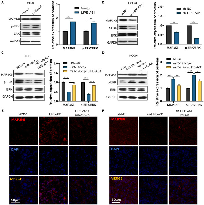
Figure 7.
The effect of LIPE-AS1/miR-195-5p on the MAPK/ERK pathway. LIPE-AS1 over- and low-expressing cell models were constructed in HeLa (A) and HCC94 cells (B), respectively. Western blot was used to measure MAP3K8 and ERK protein expression in the cells. (C,D) Transfection of cervical cancer cells with miR-195-5p mimics and inhibitors, LIPE-AS1 over-expressed plasmid or sh-LIPE-AS1. Western blot was taken to measure the protein expressions of MAP3K8 and ERK in cells. (E,F) Tissue immunofluorescence was carried out to detect MAP3K8 in the tumor tissues (the grouping was the same as Figure 5). *P < 0.05, **P < 0.01, ***P < 0.001. N = 3.

