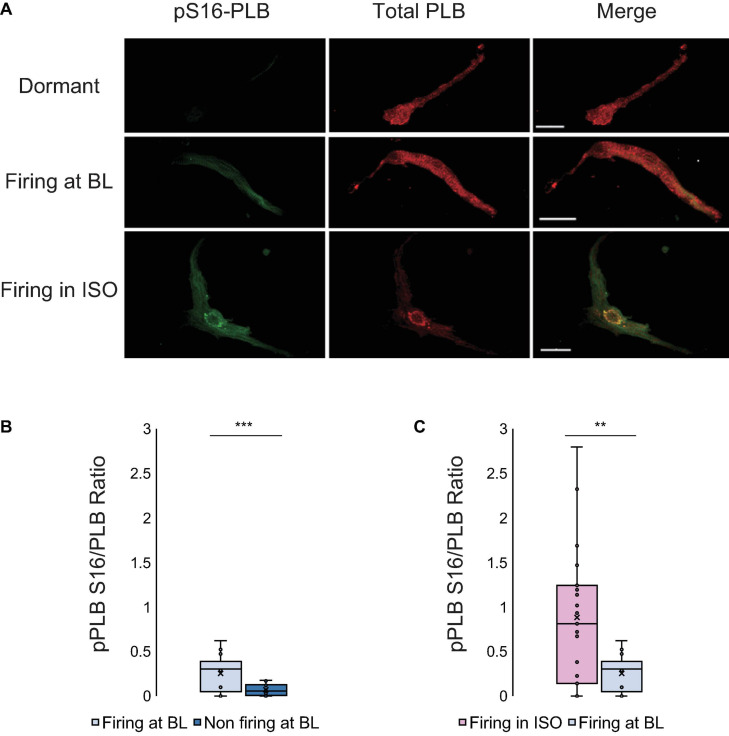
FIGURE 2.
(A) Double immunolabeling of phosphorylated phospholamban (p-PLB) at Serine 16 (green) and total PLB (red) and the merged images of SANC that were dormant at baseline (top), firing at baseline (middle) and dormant at baseline but began firing in response to isoproterenol (bottom). (B) The p-PLB/total PLB ratio of SANC firing at baseline (N = 3/n = 10) vs dormant at baseline (N = 3/n = 10) (At baseline, firing cells have much higher pPLB S16/PLB ratio than non-firing cells)] and (C) The p-PLB/total PLB ratio of SANC firing in isoproterenol (N = 5/n = 15) vs firing at baseline (N = 3/n = 10) (Firing cells in ISO have higher pPLB S16/PLB ratios than firing cells at BL). t-test, ***p < 0.001, **p < 0.01.

