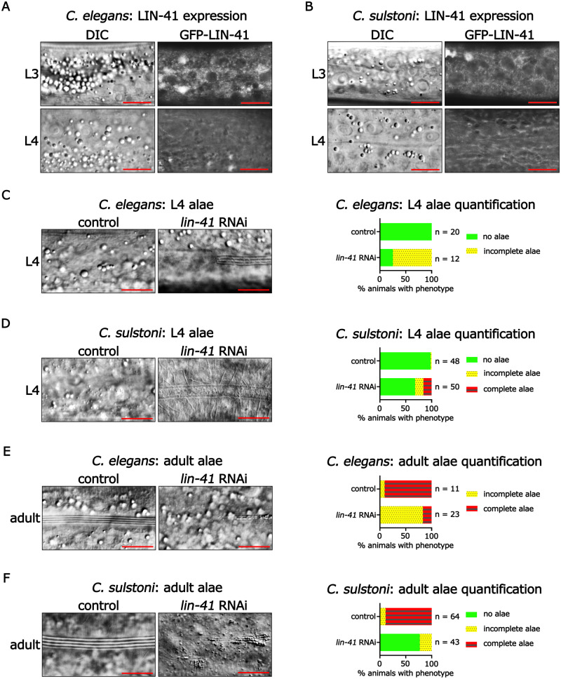
Figure 7.
lin-41 expression and function are conserved between C. elegans and C. sulstoni. (A, B) Representative DIC images (left panels) and endogenously tagged GFP-LIN-41 images (right panels) of hypodermal cells in C. elegans (A) and C. sulstoni (B) L3 (top panels) and L4 animals (bottom panels). Scale bars = 10 µM. Representative DIC images of C. elegans L4 hypodermis (C), C. sulstoni L4 hypodermis (D), C. elegans adult hypodermis (E), and C. sulstoni adult hypodermis (F) of animals fed control (empty vector) (left panels) and lin-41 RNAi (middle panels) and quantification of alae phenotypes (graphs). Scale bars = 10 µM. Note: RNAi experiments used for Figures 4, B and D and 7, D and F were performed together. Therefore, control RNAi data used for Figure 4, B and D were also used for Figure 7, D and F.

