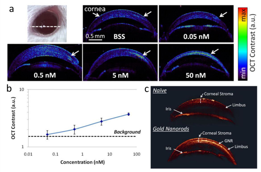
Figure 5.
Gold nanorods increased corneal OCT contrast. (a) OCT images of cross-sectional slide (white dashed line on the eye photo) through mice corneas injected with 5 μL of gold nanorods at various concentrations from 50 nM to 0 nM. (b) Quantification of the OCT contrast was observed to increase with the GNR concentration, with the lowest detectable concentration being 0.5 nM. The error bars represent standard deviation of the OCT signal. (c) 3D rendering of OCT images show a much brighter OCT signal from the corneal stroma injected with 10 μL of GNR at 50 nM (lower eye) than naïve corneal stroma (upper eye). The corneal structures (corneal stroma, limbus and iris) exhibit very consistent OCT signal intensities across both the naïve and injected eyes. Reproduced from ref. 96 with permission from John Wiley and Sons, copyright [2020].

