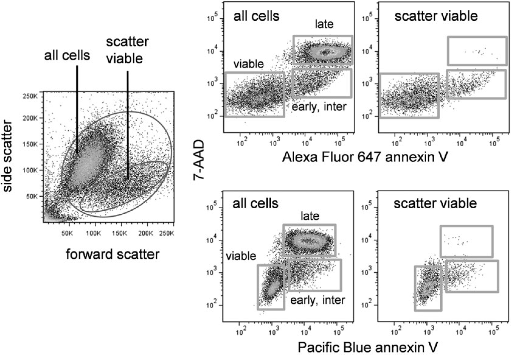
Fig. 3.

Annexin V and DNA dye labeling in apoptotic cells. EL4 cells were treated with camptothecin at 5 μM for 16 h. Left dotplot shows forward versus side scatter of treated cells. Top right row shows Alexa Fluor 647 annexin V versus 7-AAD labeling for all cells (left plot) or scatter viable cells only (right plot). Bottom right row shows Pacific Blue annexin V versus 7-AAD labeling for all cells (left plot) or scatter viable cells only (right plot)
