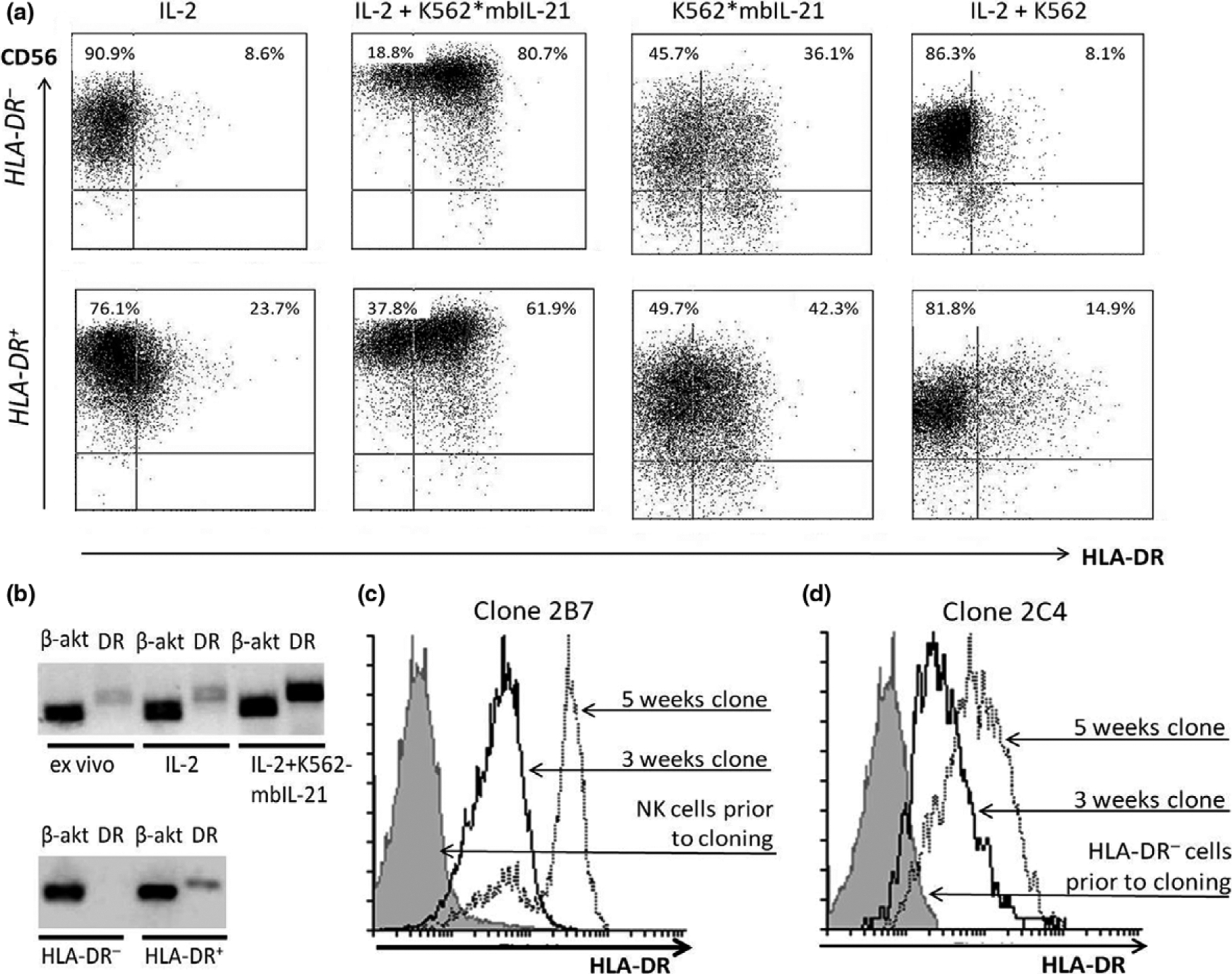
Figure 4.

NK cell stimulation with IL-2 and/or K562-mbIL21 triggers HLA-DR surface expression on HLA-DR-negative cells. (a) HLA-DR expression in HLA-DR+ and HLA-DR− NK cell subsets after 6 days of incubation with indicated stimuli. (b) Presence of HLA-DR α-subunit mRNA in NK cells, freshly isolated and after 6 days of stimulation (upper row), and in HLA-DR-positive and HLA-DR-negative NK cells directly after sorting in “Single cell” mode (bottom row). (a, b) Representative data obtained from a single donor out of three donors examined are shown. (c) Changes in HLA-DR expression in clones, obtained from freshly isolated unsorted NK cells. (d) Changes in HLA-DR expression in clones, obtained from freshly isolated HLA-DR-negative NK cells subset. Representative data obtained from a single clone out of 20 (c) or 24 (d) clones examined, respectively, are shown in each figure.
