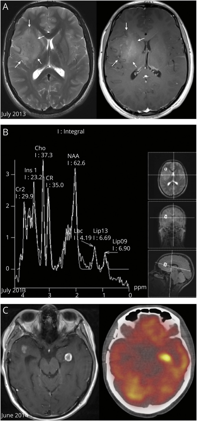
Figure 1. Radiologic Features of the Brain Tumors.

(A) Axial planes of T2 turbo spin echo-weighted brain MRI (left image) and T1 gadolinium sequence (right image) showing a right fronto-insular lesion with high signal on T2 (arrows) and with inhomogeneous enhancement on T1, corresponding to a high-grade lesion. (B) Magnetic resonance spectroscopy (MRS) shows a raised choline peak and N-acetyl aspartate peak (resonating at 3.7 and 6.2 ppm, respectively), suggestive of anaplastic glioma. (C) Gadolinium T1-weighted brain MRI (left image) showing tumor relapse with 2 new enhancing left temporal lesions (the left one was biopsied stereotactically) and the corresponding hypermetabolic image on PET CT (right).
