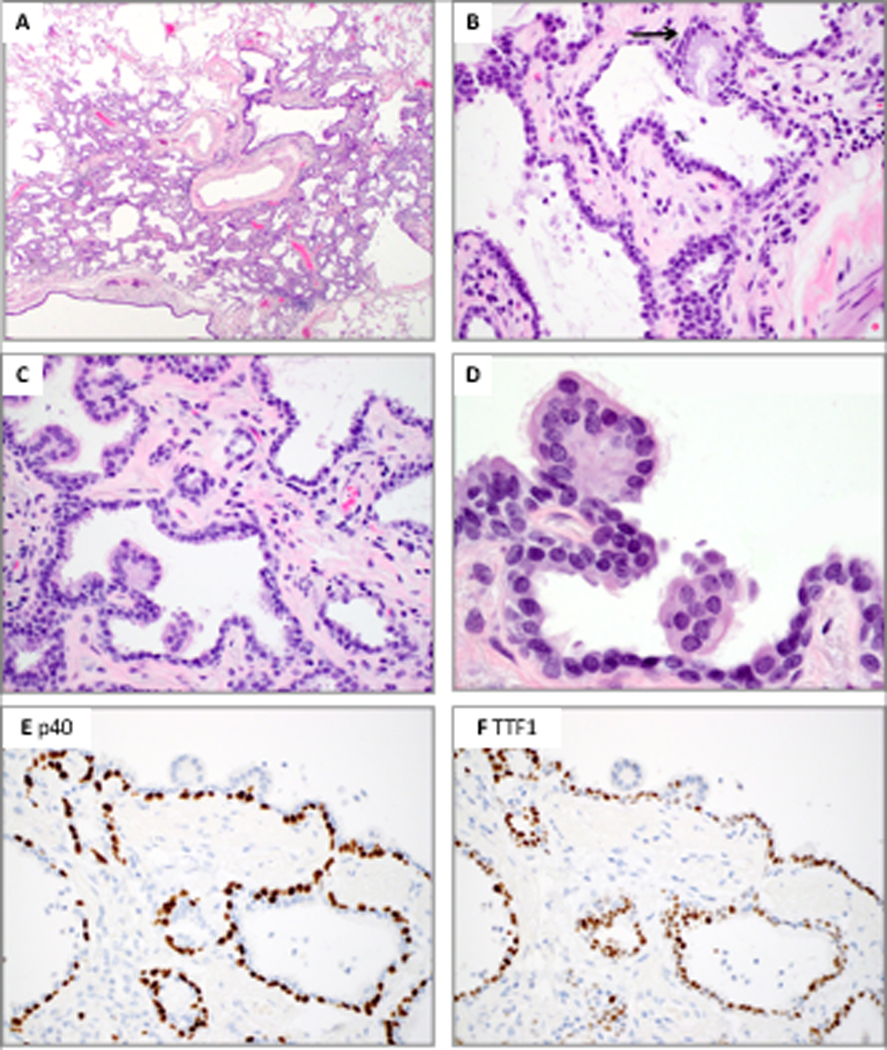
Figure 3.

Distal-type bronchiolar adenoma with entirely flat architecture and focal mucin and cilia (Patient 9, case 2). A, Low power shows peribronchiolar location penetrated by medium-sized muscular arteries. B, Mucinous cells are only present in rare glandular crypts (arrow). C and D, Ciliated cells are inconspicuous and only observed on top of micropapillary-like tufts. The luminal cells show predominance of cuboidal cells that resemble type II pneumocytes. E, P40 highlights a continuous basal cell layer. F, TTF1 is weakly positive in the basal cells and moderately to strongly positive in non-ciliated luminal cuboidal cells; ciliated micropapillary tufts are negative.
