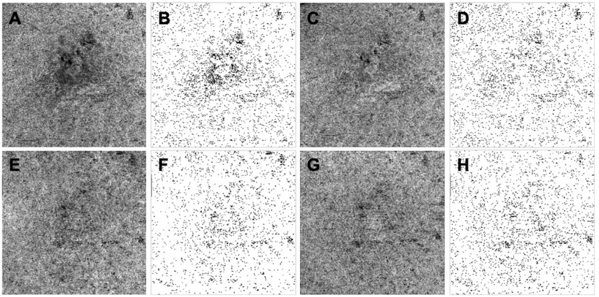
Figure 4:

Phansalkar threshold method applied to uncompensated and compensated choriocapillaris (CC) images before and after drusen collapse. A, B, C, D: images before drusen collapse; E, F, G, H: images after drusen collapse; A, E: Uncompensated CC en face flow images using a custom slab with a thickness of 15 located 16 microns below Bruch’s membrane; B, F: CC binary maps thresholded using the Phansalkar method with a 3 pixel window radius based on images A and E; C, G: Compensated CC en face flow images before and after the drusen resolved using a validated compensation strategy; D, H: CC binary maps thresholded using the Phansalkar method with a 3 pixel window radius based on images C and G.
