Figure 1.
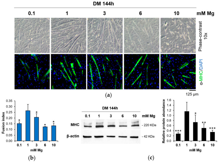
Low and high Mg concentrations inhibit myogenesis. C2C12 cells were cultured in differentiation medium (DM) for 144 h in the presence of different extracellular concentrations of Mg. (a) Pictures were taken with optical microscope (10× magnification, upper panels). After immunofluorescence with antibodies against Myosin Heavy Chain (MHC; green fluorescence), images were acquired using a fluorescence microscope (10× magnification, lower panels). The nuclei were stained with 4′,6-diamidino-2-phenylindole (DAPI). (b) Fusion index was calculated as the ratio of the number of nuclei within myotubes (>2 nuclei) to the total number of nuclei in the field and quantified based on (a). (c) MHC levels were analyzed by Western blot. β-actin was used as control of loading. A representative blot (left) and densitometry performed on three independent experiments and obtained by ImageLab (right) are shown. * Indicates significance with respect to 1 mM Mg (* p ≤ 0.05; ** p ≤ 0.01; *** p ≤ 0.001).

