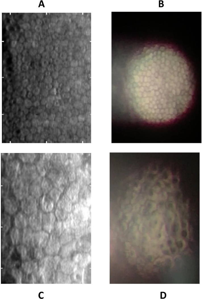
Figure 1.

Comparison of Konan specular microscope to smartphone imaging. In a healthy cornea (A), standard specular microscopy reveals characteristic hexagon-shaped endothelial cells. Smartphone specular microscopy (B) demonstrates a circular field of view, with reflective artifact of specular reflection to the left. Qualitative analysis of pathology is seen in a case of posterior polymorphous corneal dystrophy visualized using the standard specular microscope (C), which reveals large endothelial cells and a few apparent opacities. Smartphone specular microscopy highlights intracellular opacities not seen in images of normal corneas.
