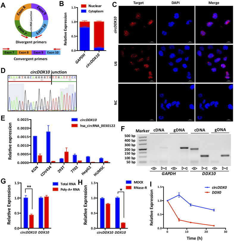
Figure 1.
Subcellular localization, expression levels and biological properties of circDDX10 in granulosa cells (GCs). (A) Schematics of back-to-back and face-to-face primers (black vertical line indicates the back-splicing site; red arrow represents the amplification direction of the upstream and downstream primers). (B) CircDDX10 nucleoplasm distribution in COV434 (granulosa cell line). (C) Subcellular localization of circDDX10 in COV434 using FISH technique. (U6 was used as a positive control for nuclear. NC, negative control. Bar = 20 μm). (D) Sanger sequencing of circDDX10. (Black vertical line indicates the back-splicing site. Red horizontal line representing 3' end and green horizontal line represents 5' end). (E) The relative expression levels of circDDX10 in different cell lines (hsa_circRNA_0030122 was used as control). (F) Properties of back-splicing. (gDNA and cDNA were used as templates. And back-to-back primers and face-to-face primers were used for amplification. GAPDH as an internal control). (G) poly-A- RNA characterization. (Total RNA and poly-A+ RNA were used for reverse transcription into cDNA, respectively). (H) Properties of resistance to RNase R digestion. (I) Properties of stability. All experiments were repeated for three times and the data were expressed as mean ± standard deviation. *, P < 0.05; **, P < 0.01.

