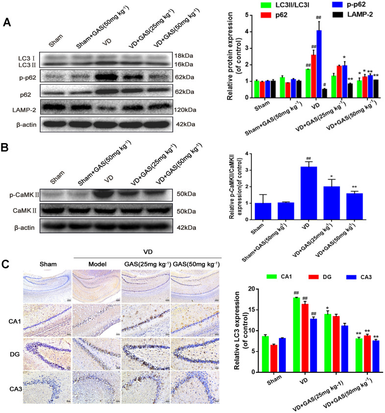
Figure 2.
GAS reversed the suppression of autophagy flux and hyperphosphorylation of CaMKII in VD rats. (A, B) The protein extract of hippocampal tissue was analyzed by western blotting for LC3, p62, p-p62 (Thr349), LAMP-2, CaMKII, and p-CaMKIIα (Thr286). Protein levels were quantified and normalized to β-actin. Data are presented as the mean ± standard error of the mean (SEM). ##P< 0.01 versus sham, *P<0.05, **P< 0.01 versus model. (C) Representative images of hippocampal tissue sections immunostained with LC3 antibodies (×50, ×200). Scale bars: 200 μm or 50 μm. LC3, microtubule-associated protein 1 light chain 3; LAMP-2, lysosomal-associated membrane protein-2; CaMKII, Ca2+-calmodulin stimulated protein kinase II; p-CaMKIIα, phosphorylated CaMKIIα.

