
Figure 7.
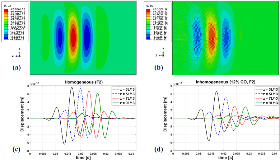
Top row: A snapshot in time of the propagating waveform in the FE simulation for (a) the homogeneous medium (b): the inhomogeneous medium. Displacement amplitudes will scale with source excitation in this linear model. Bottom row: Time evolution of shear waves at four different locations along a single line in the z-direction in the FE simulations in the (c) homogeneous and (d) inhomogeneous medium. All cases are at the fibrosis (stiffness) level of F4. The inhomogeneous medium has 12% inclusions.