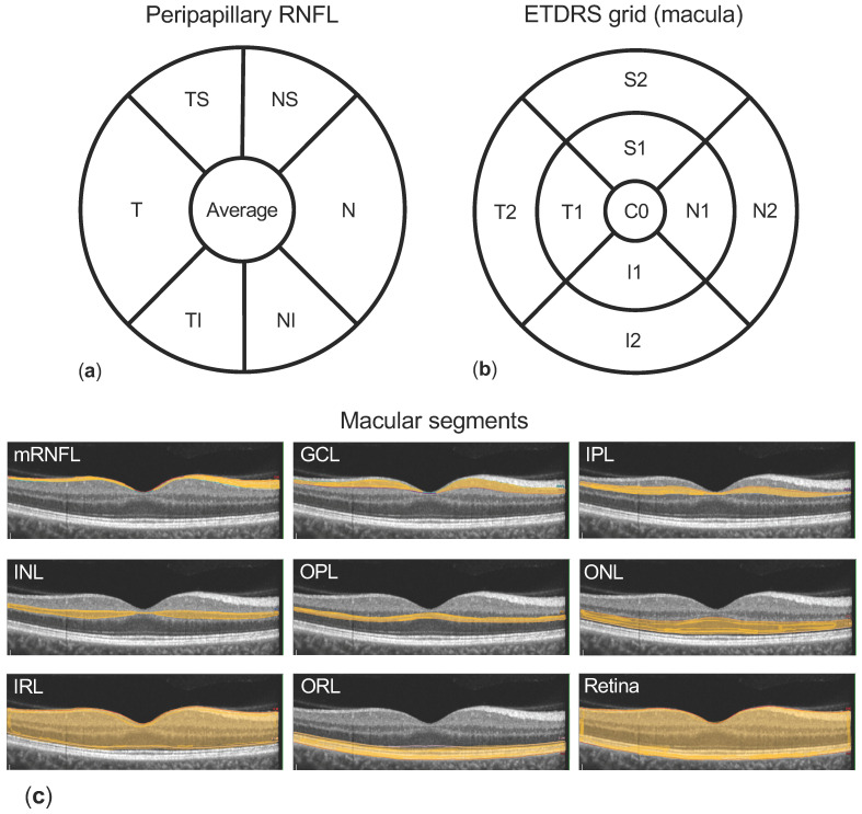
Figure 1.
Methodology. (a) shows the peripapillary retinal nerve fibre layer (pRNFL) quadrants and sectors (T: temporal, N: nasal, superior separated into TS: temporal superior and NS: nasal superior, and inferior separated into TI: temporal inferior and NI nasal inferior) measured by the optical coherence tomography (OCT) software. Macular segments (c) are separated semi-automatically (macular retinal nerve fibre layer (mRNFL), ganglion cell layer (GCL), inner plexiform layer (IPL), inner nuclear layer (INL), outer plexiform layer (OPL), outer nuclear layer (ONL)–grouped as inner retinal layers (IRL), retinal pigment epithelium (RPE), and outer retinal layers (ORL)), and the thickness of each layer is reported using the Early Treatment Diabetic Retinopathy Study (ETDRS) grid 1, 3.5, 6 mm (b) containing nine subfields (C0: centre, S1 and S2 superior, N1 and N2 nasal, I1 and I2 inferior, and T1 and T2 temporal).

