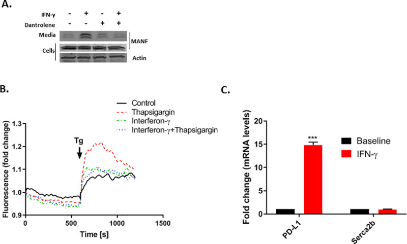
Fig 4. MANF release in response to IFN-γ treatment is mediated by ER Ca2+ depletion.

(A) SKMEL28 cells were treated with 100 ng/ml IFN-γ and/or 30μM Dantrolene as indicated for 48 hours, followed by immunoblot analysis of the cells and media. Data are representative of three independent experiments. (B) SKMEL28 cells were treated for 48 hours with IFN-γ or control as indicated prior to loading with Fluo-3-AM. Thereafter, the medium was switched to 1mM EGTA in HBS and supplemented after 10 min with 1 μM Tg (indicated by the arrow) for the Thapsigargin and IFN-γ+Thapsigargin treated cells (red or blue dotted lines, respectively), to release any Calcium remaining in the ER, while DMSO was added to the control and IFN-γ (only) treated cells (black and green dotted lines, respectively). Increase in cytosolic calcium was monitored by fluorescence tracing. One representative experiment of four is shown. (C) Skmel28 cells were treated with 100 ng/ml IFN-γ for 48 hours, followed by RT-PCR analysis of SERCA2b and PD-L1 (lower panels). RNA expression is relative to baseline. N = 3, data is represented as mean ± SEM. *** p-value<0.001.
