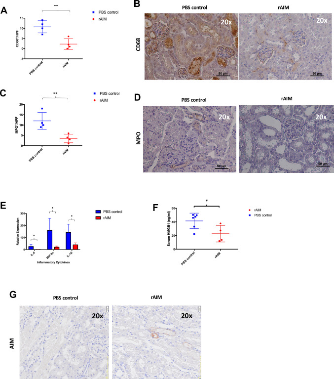
Fig 2. Recombinant AIM administration alleviates local tissue and systemic inflammation following syngeneic renal transplantation.
(A-D) Kidney tissue sections were stained for CD68 or MPO to detect graft-infiltrating macrophages and granulocytes, respectively. (A) Number of CD68+ macrophages/HPF. (B) Immunohistochemistry images of kidney graft sections staining CD68. (C) Number of MPO+ granulocytes/HPF. (D) Immunohistochemistry images of kidney graft sections staining MPO. (E) Measurement of pro-inflammatory cytokines (IL-6, MIP-2ɑ, and IL-1β) using quantitative RT-PCR. Data were normalized to GAPDH gene expression. *p<0.05, **p<0.01, n = 4/group. (F) Serum HMGB1 levels were quantified using ELISA. p = 0.1, n = 4-6/group. (G) Kidney tissue sections were stained for AIM.

