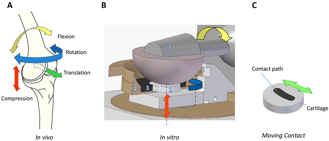
Figure 1. Physiologic joint kinematics and bioreactor set up.

A) Representative primary and secondary motions of the knee joint (in vivo) and compression (red arrow); flexion and extension (yellow arrow); internal and external rotation (blue arrow); anterior and posterior translation (green arrow); medial/lateral translation and abduction/adduction movement not shown. B) Bioreactor set up for in vitro testing. Ceramic ball [1] provides compression (red arrow) and articulation (yellow arrow) that translate into shear forces at the explant surface (2 /grey disk). Off-center axis rotation (blue arrow) simulates secondary motions (translation and rotation). Cartilage explant is held within a porous polyethylene scaffold (3), which is held in place by a dished PEEK cup holding the testing media. Representative image shows a horizontal cross section through the ceramic ball (1) and a vertical cross section through the porous scaffold (3) and PEEK cup (4). C) Migrating contact /MC articulation: Ceramic ball moves along curvilinear contact path (black) on the cartilage explant. In SC articulation, forces and motions are confined to a circular region within the center of the cartilage explant (not shown).
