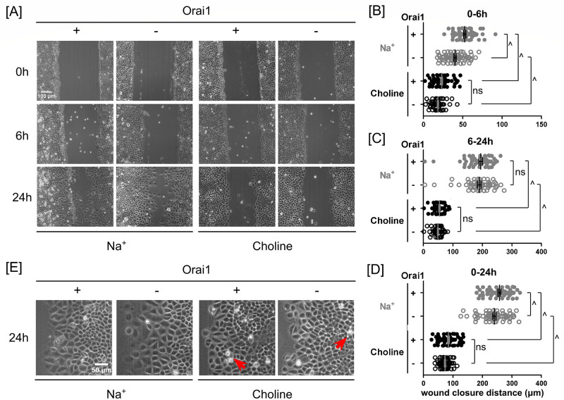
Fig 6. In the presence of extracellular Na+, Orai1 silencing slowed endothelial wound closure.
(A) Phase-contrast images of scratched Orai1-expressing and -silenced PA2879 endothelial monolayers at 0, 6 and 24 hours in the presence and absence of extracellular Na+. Scatter plots of wound closure distance in (B) 0–6 hours, (C) 6–24 hours, and (D) 0–24 hours. In the presence of extracellular Na+, wound closure distances in Orai1-silenced PA2979 monolayers were less than the distances in Orai1-expressing monolayers. However, in the absence of extracellular Na+, the contribution of Orai1 was not significant. Data were reported as mean ± SEM. (E) Magnified view of the marginal regions of the Orai1-expressing and -silenced cells at 24 hours of wound closure in Na+- and choline-containing media. Cells near the wound edge spread larger in Na+- than in choline-containing media. Intercellular gaps were observed in the absence of extracellular Na+. Arrows point to typical interendothelial gaps. Statistical significance was assessed using one-way ANOVA with Tukey’s post hoc test (ns—not significant, ^—P < 0.0001).

