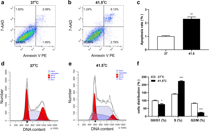
Fig. 3.
The effect of HS on proliferation of 3T3-L1 preadipocytes was investigated by detecting cell apoptosis and cell cycle. a-c HS induced apoptosis of 3T3-L1 preadipocytes. The cells were exposed to 37 °C or 41.5 °C for 2 d, analyzed by flow cytometry. In the two-dimensional FCM analysis, the cells were divided into 4 subgroups: the lower left quadrant was the living cell group, the lower right quadrant was the early apoptotic cells, the upper right quadrant was the late apoptotic cells, and the upper left quadrant was the necrotic cells. d-f HS induced S phase arrest of 3T3-L1 preadipocytes. The cells were exposed to 37 °C or 41.5 °C for 2 d, analyzed by flow cytometry. The data represent the mean ± SEM (n = 3). * The mean difference was significant (P < 0.05) and ** or *** mean difference were extremely significant (P < 0.01, P < 0.001), compared with 37 °C

